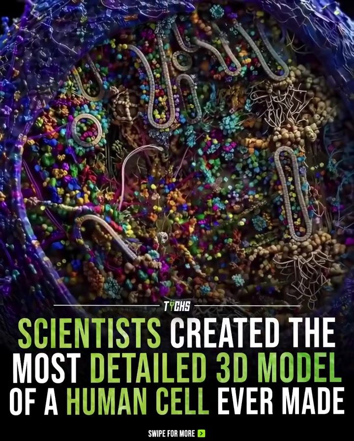
3D Model of a Human Cell
The 3D model of a human cell is a groundbreaking scientific visualization that offers an unprecedented look into the intricate world of cellular structures. This detailed model wasn't captured by a camera but was meticulously created using a combination of advanced experimental data and cutting-edge techniques. Each molecule in this eukaryotic cell was individually modeled using real data from methods such as X-ray crystallography, nuclear magnetic resonance (NMR), and cryo-electron microscopy.
Context / Why This Matters
Understanding the complex structures within a cell is crucial for both scientific research and educational purposes. The 3D model provides an unparalleled level of detail, showing thousands of molecular structures working together. This visualization helps scientists and students grasp the intricate processes that power every living organism.
Main Discussion
The Creation Process
The creation of this 3D model involved several sophisticated techniques:
-
X-ray Crystallography: This method involves directing X-rays at a crystal of the molecule being studied, which then diffracts in a pattern that can be analyzed to determine the atomic and molecular structure. It's particularly useful for studying protein structures.
-
Nuclear Magnetic Resonance (NMR): This technique uses a magnetic field and radio waves to observe the local magnetic field around atomic nuclei. It is valuable for studying the structure and dynamics of molecules in solution, making it ideal for understanding proteins and nucleic acids.
-
Cryo-Electron Microscopy (Cryo-EM): This method involves flash-freezing samples and then using electron beams to create an image. It's particularly useful for studying large molecular complexes and viruses.
The Result
By combining data from these techniques, scientists were able to create a single, incredibly detailed cross-section of a cell. This model visualizes thousands of molecular structures working together, providing a level of detail that no camera could capture. The result is a vibrant and colorful 3D model that showcases the intricate structures within a cell.
The Importance of Visualizations
Visualizations like this are essential for several reasons:
-
Educational Tool: For students, these models provide a clear and detailed understanding of cellular structures and processes. They can see how different molecules interact and function within the cell, making complex concepts easier to grasp.
-
Research Tool: For scientists, these visualizations offer new insights into molecular structures and their functions. They can use these models to study diseases at a molecular level, develop new treatments, and understand how different molecules interact.
Practical Tips
If you are interested in exploring 3D cellular models, here are some tips to get started:
-
Access Online Resources: Many educational institutions and scientific organizations offer online resources and interactive 3D models. Websites like the Protein Data Bank (PDB) and the National Center for Biotechnology Information (NCBI) provide detailed information and visualizations of molecular structures.
-
Use Software Tools: There are several software tools available for creating and viewing 3D models. PyMOL and Chimera are popular choices for visualizing molecular structures. They allow you to manipulate and analyze the models in great detail.
-
Attend Workshops and Seminars: Many universities and research institutes offer workshops and seminars on molecular visualization. These can provide hands-on experience and in-depth knowledge about the techniques used to create these models.
Important Takeaways
-
Advanced Techniques: The 3D model of a human cell was created using advanced techniques like X-ray crystallography, NMR, and cryo-EM, providing an unprecedented level of detail.
-
Educational and Research Value: These visualizations are invaluable for both educational and research purposes, offering new insights into cellular structures and functions.
-
Accessibility: There are many online resources and software tools available for exploring 3D cellular models. These can be a great starting point for anyone interested in learning more about cellular structures.
Conclusion
The 3D model of a human cell is a testament to the power of scientific visualization. By combining experimental data from various techniques, scientists have created a detailed and vibrant representation of cellular structures. This model not only helps in understanding the intricate processes within a cell but also serves as a valuable tool for both education and research. Whether you're a student or a scientist, exploring these 3D models can offer a deeper understanding of the hidden world that powers every living organism.
Questions readers ask
What makes the 3D model of a human cell so significant?
The 3D model of a human cell is significant because it offers an unprecedented level of detail, showing thousands of molecular structures and their interactions. This level of detail is crucial for understanding complex cellular processes and advancing scientific research.
How is the 3D model of a human cell created?
The 3D model is created using a combination of advanced experimental data and cutting-edge techniques. Methods such as X-ray crystallography, nuclear magnetic resonance (NMR), and cryo-electron microscopy are employed to gather real data, which is then used to individually model each molecule in the cell.
What are the benefits of using 3D models in cell research?
3D models provide a unique perspective on human cells, making it easier to visualize intricate structures and molecular interactions. They serve as powerful tools for understanding complex cellular processes and can reveal details that would be difficult to observe through traditional microscopic methods.
What techniques are used to visualize the detailed structure of a human cell?
Several advanced techniques are used to visualize the detailed structure of a human cell, including X-ray crystallography, nuclear magnetic resonance (NMR), and cryo-electron microscopy. These methods provide the necessary data to create accurate and detailed 3D models of cellular structures.
How does the 3D model help in understanding complex cellular processes?
The 3D model provides an unparalleled level of detail, showing thousands of molecular structures working together. By visualizing these interactions, scientists can gain a deeper understanding of how different parts of the cell function and interact, which is essential for advancing research in various fields.
What is the difference between a 3D cell model and traditional microscopic images?
Traditional microscopic images provide a 2D representation of cellular structures, while 3D cell models offer a detailed, three-dimensional view. This allows for a more comprehensive understanding of molecular interactions and cellular processes, as it provides a more accurate representation of the cell's complex structure.
Who can access and benefit from these 3D cell models?
3D cell models are primarily used by researchers and scientists in various fields, such as biology, biochemistry, and medicine. These models can also be utilized by medical professionals, providing a more detailed understanding of cellular structures and processes, which can aid in diagnosing and treating diseases.
Related deep dives
Similar reads based on topic and creator.




Recent articles
Fresh deep dives from the latest Reels we unpacked.




Comments
Be the first to comment.