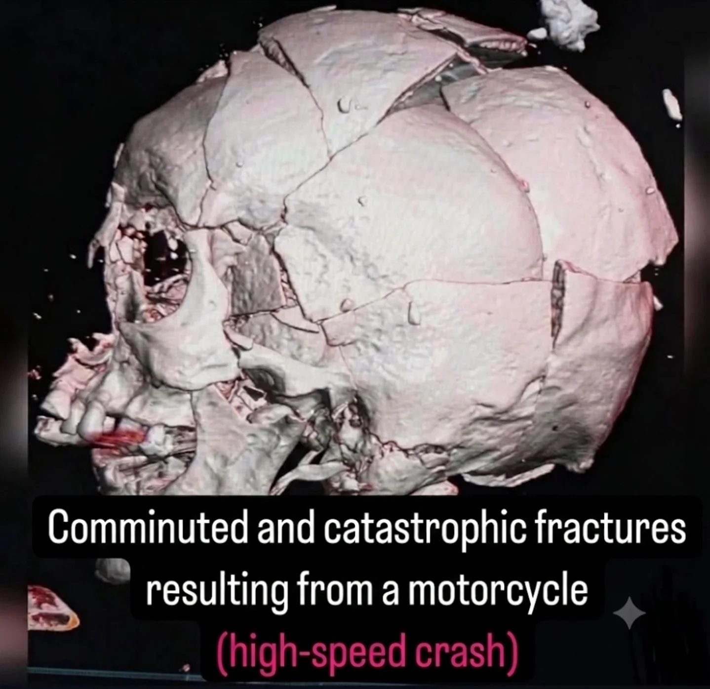
Medical Imaging of Skull Fractures
When dealing with severe trauma, medical imaging plays a critical role in assessing and diagnosing injuries. One of the most severe types of trauma involves comminuted and catastrophic fractures of the skull, often resulting from high-velocity impacts such as high-speed motorcycle crashes. These injuries present significant challenges for medical professionals and require immediate, multidisciplinary intervention.
Context
Severe craniomaxillofacial trauma can lead to extensive fractures of the neurocranium and viscerocranium. The neurocranium protects the brain, while the viscerocranium encompasses the facial bones. Injuries to these areas can be life-threatening and require prompt medical attention. High-velocity collisions, such as those involving motorcycles, transfer immense kinetic energy to the head, leading to complex and devastating fractures.
Injuries Explained
Comminuted and Catastrophic Fractures
Comminuted fractures occur when the bone shatters into multiple fragments. These fractures are often the result of high-impact forces and are particularly challenging to treat. Catastrophic fractures are a severe form of comminuted fractures, often resulting in extensive fragmentation and displacement of bone pieces. In the context of the skull, these fractures can compromise the cranial vault, leading to a "cracked eggshell" appearance.
Affected Bones
Severe skull fractures often involve multiple bones, including the frontal, parietal, and temporal bones. These bones are critical for protecting the brain and maintaining the structural integrity of the skull. When these bones are fractured, the risk of traumatic brain injury (TBI) increases significantly.
Structural Compromise
The fractures extend across these bones, resulting in a compromised cranial vault. This structural compromise can lead to further complications, including intracranial pressure issues and potential brain herniation. Stabilizing the skull and managing intracranial pressure are crucial steps in the initial treatment process.
The Role of Medical Imaging
3D Computed Tomography (CT) Reconstruction
3D computed tomography (CT) scans are invaluable in diagnosing and assessing the extent of skull fractures. These scans provide detailed, three-dimensional images of the skull, allowing medical professionals to visualize the fractures from multiple angles. This level of detail is essential for planning surgical interventions and understanding the full extent of the damage.
Visualizing Fractures
The 3D CT reconstruction reveals multiple displaced and fragmented fracture lines. These lines extend across various bones, providing a clear picture of the structural compromise. The visualization helps in identifying the exact locations and severity of the fractures, guiding the surgical team in their approach to reconstruction.
Clinical Presentation
In a clinical setting, the severity of these injuries presents immediate challenges. Medical teams must act quickly to stabilize the patient, manage potential brain injuries, and reconstruct the craniofacial structure. This often involves a multidisciplinary approach, including neurosurgery and maxillofacial surgery.
Practical Tips
Emergency Response
In cases of high-velocity trauma, immediate stabilization is paramount. This includes securing the patient’s airway, breathing, and circulation (ABCs), as well as controlling any bleeding and immobilizing the spine. Rapid transportation to a trauma center with specialized medical imaging capabilities is crucial.
Diagnostic Imaging
Once the patient is stable, 3D CT scans should be performed to assess the extent of the fractures. These scans provide detailed information that guides the surgical plan. It’s essential to have access to advanced imaging technology and expertise in interpreting the results.
Surgical Intervention
Surgical intervention often involves multiple steps, including:
- Stabilizing Intracranial Pressure: This may require placement of an intracranial pressure (ICP) monitor and administration of medications to reduce swelling.
- Managing Traumatic Brain Injury (TBI): Careful monitoring and treatment of potential brain injuries are critical.
- Reconstructing the Cranial Vault: This involves meticulous surgical procedures to realign and stabilize the fractured bones, often using plates, screws, and bone grafts.
Important Takeaways
- High-velocity Trauma: Such injuries are often characterized by comminuted and catastrophic fractures, especially in high-speed motorcycle crashes.
- Medical Imaging: 3D CT scans are essential for diagnosing and planning treatment for severe skull fractures.
- Multidisciplinary Care: Effective treatment requires a team approach involving neurosurgery, maxillofacial surgery, and other specialists.
- Immediate Action: Rapid stabilization and transfer to a specialized trauma center are crucial for patient survival and outcomes.
Conclusion
Severe skull fractures resulting from high-impact trauma present significant challenges for medical professionals. The use of advanced medical imaging tools, such as 3D CT scans, is essential for accurate diagnosis and effective treatment planning. Understanding the extent and nature of these injuries helps in developing a comprehensive approach to managing and reconstructing the damaged skull. With timely and specialized intervention, patients have a better chance of recovery and rehabilitation.
Questions readers ask
What are comminuted and catastrophic skull fractures?
Comminuted skull fractures involve the bone breaking into multiple pieces, while catastrophic fractures are severe and often life-threatening, affecting multiple skull bones. These types of fractures are commonly seen in high-speed motorcycle crashes due to the significant impact forces involved.
Why is 3D medical imaging important for assessing severe skull fractures?
3D medical imaging, such as CT scans, provides detailed, three-dimensional information about the extent and complexity of skull fractures. This helps medical professionals to plan surgical interventions accurately and assess the impact on surrounding structures like the brain and facial bones.
What are the immediate concerns when treating severe skull fractures from motorcycle crashes?
Immediate concerns include stopping any bleeding, stabilizing the patient, and preventing further damage to the brain. Severe skull fractures can lead to brain herniation, hemorrhage, and other life-threatening complications, making timely intervention crucial.
How does severe craniomaxillofacial trauma affect the body?
Severe craniomaxillofacial trauma can involve extensive fractures of both the neurocranium, which protects the brain, and the viscerocranium, which includes facial bones. These injuries can compromise vital functions, such as breathing and vision, and often require immediate, multidisciplinary intervention.
What role do CT scans play in diagnosing skull fractures?
CT scans are essential for diagnosing skull fractures, particularly in high-velocity impacts like motorcycle crashes. They offer clear, detailed images that help identify the exact location, type, and severity of fractures, guiding doctors in formulating an appropriate treatment plan.
Can you explain the importance of a multidisciplinary approach in treating motorcycle crash injuries?
A multidisciplinary approach is vital because motorcycle crash injuries can be complex and affect multiple body systems. This approach brings together specialists from various fields, such as neurosurgery, orthopedics, and maxillofacial surgery, to provide comprehensive care and improve patient outcomes.
Related deep dives
Similar reads based on topic and creator.



Recent articles
Fresh deep dives from the latest Reels we unpacked.




Comments
Be the first to comment.