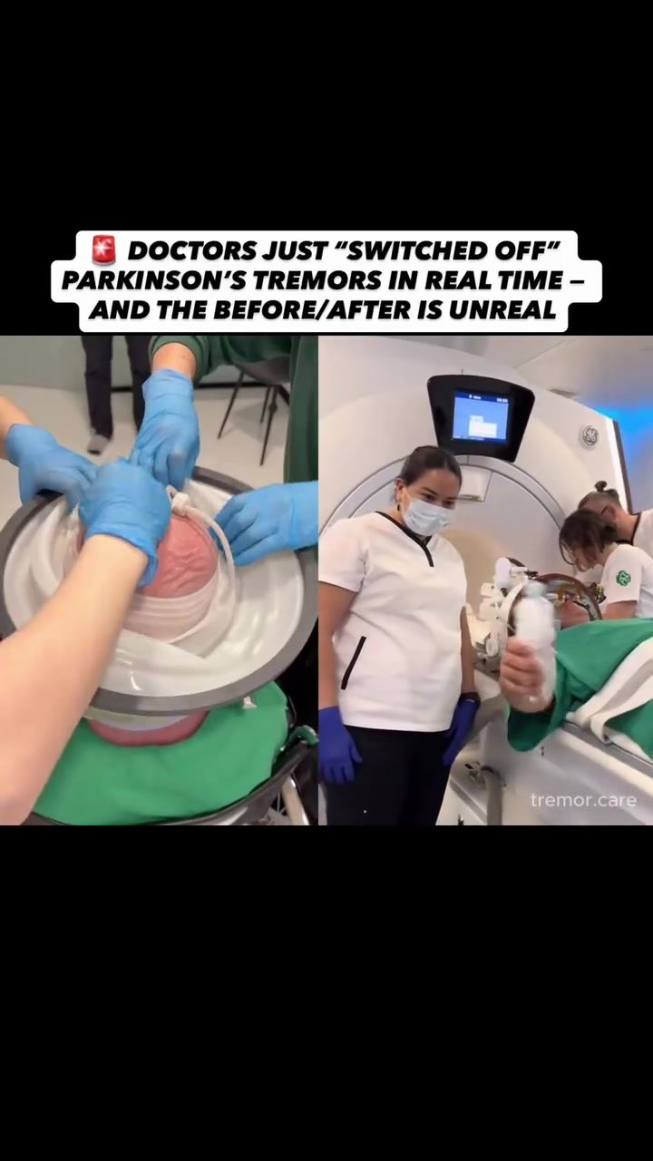
MR-Guided Focused Ultrasound for Parkinson's Tremors
Parkinson's disease is a progressive neurological disorder that affects millions of people worldwide. One of the most debilitating symptoms is tremor, an involuntary shaking that can severely impact a patient's quality of life. Recent advancements in medical technology offer new hope for managing these tremors effectively. MR-guided focused ultrasound is one such innovation that has shown promising results in reducing or even eliminating Parkinson's tremors in real-time.
Context / Why This Matters
Tremors in Parkinson's disease can be particularly challenging to manage. Traditional treatments often involve medications that may have side effects or invasive procedures like deep brain stimulation, which requires surgery and implantation of electrodes. The introduction of MR-guided focused ultrasound provides a non-invasive alternative that targets specific areas of the brain responsible for tremors, offering a more precise and less invasive treatment option.
The Role of MR-Guided Focused Ultrasound
Understanding the Technology
MR-guided focused ultrasound combines the power of magnetic resonance imaging (MRI) with ultrasound technology. The MRI provides real-time imaging, allowing doctors to precisely target the area of the brain causing the tremors. High-intensity focused ultrasound waves are then directed to this specific area, creating heat that disrupts the abnormal neural activity responsible for the tremors. This process is non-invasive, meaning it does not require any open surgery or implants.
How It Works
The procedure involves the patient being positioned inside an MRI machine, where the ultrasound waves are precisely directed to a tiny area deep in the brain. The MRI provides continuous feedback, ensuring that the ultrasound waves are accurately targeted. This real-time guidance is crucial for the success of the procedure, as it allows doctors to make immediate adjustments if necessary.
Immediate Results
One of the most striking aspects of this technology is the immediate results it can achieve. Within moments of the procedure, patients who were previously experiencing uncontrollable shaking can suddenly regain control of their movements. The split-screen comparisons of before and after effects highlight a significant reduction in tremors, demonstrating the effectiveness of this approach.
Practical Tips for Patients and Caregivers
What to Expect
If you or a loved one is considering MR-guided focused ultrasound for Parkinson's tremors, it's essential to understand what the procedure entails. The process is typically outpatient, meaning patients can go home the same day. There is no recovery time required as there would be with open surgery. However, it's important to discuss the potential benefits and risks with a healthcare provider before proceeding.
Preparing for the Procedure
Preparation for MR-guided focused ultrasound involves several steps. Patients will undergo a detailed assessment to ensure they are suitable candidates for the procedure. This may include additional imaging tests and consultations with a neurologist and a radiologist. It's also important to follow any pre-procedure instructions provided by the medical team, such as fasting or avoiding certain medications.
Post-Procedure Care
Following the procedure, patients may experience some mild discomfort or fatigue, but these symptoms usually resolve quickly. It's crucial to follow up with the healthcare provider to monitor progress and address any concerns. Regular check-ups and follow-up appointments will help ensure that the treatment is working as intended and that any potential side effects are managed effectively.
Important Takeaways
MR-guided focused ultrasound represents a significant advancement in the treatment of Parkinson's tremors. Its non-invasive nature, combined with real-time imaging and precise targeting, offers a promising alternative to traditional treatments. The immediate and dramatic improvement in tremor symptoms makes it a compelling option for patients seeking relief from this debilitating condition.
Moreover, the procedure's success in providing instant results underscores its potential to improve the quality of life for Parkinson's patients. As more research and clinical trials continue, this technology may become a standard treatment option for managing tremors associated with Parkinson's disease.
Conclusion
The advancement of MR-guided focused ultrasound in treating Parkinson's tremors is a testament to the incredible potential of modern medical technology. By offering a non-invasive, precise, and effective solution, this technology provides new hope for those battling this challenging condition. As awareness and understanding of this procedure grow, it holds the promise of transforming the lives of many Parkinson's patients, offering them a chance to regain control and improve their daily functioning.
Questions readers ask
How does MR-guided focused ultrasound work to reduce Parkinson's disease tremors?
MR-guided focused ultrasound uses MRI technology for real-time imaging and ultrasound waves to target specific brain regions. The ultrasound waves disrupt abnormal neural activity, which helps to reduce or even eliminate tremors. This process is non-invasive and does not require surgery.
What are the benefits of using MR-guided focused ultrasound for Parkinson's disease tremors?
The primary benefits include being non-invasive, precision targeting of affected brain regions, real-time adjustments during the procedure, and the potential for significant tremor reduction. Unlike traditional treatments, this method does not involve medications with potential side effects or invasive surgeries.
Who is a good candidate for MR-guided focused ultrasound treatment for Parkinson's tremors?
Good candidates are typically those who have Parkinson's disease with tremors that significantly impact their daily life and have not responded well to medication. A thorough evaluation by a medical team, including a neurologist, is necessary to determine if this treatment is suitable.
How does this treatment compare to deep brain stimulation for Parkinson's disease tremors?
Unlike deep brain stimulation, which involves brain surgery and the implantation of electrodes, MR-guided focused ultrasound is non-invasive. Both methods aim to disrupt abnormal neural activity, but ultrasound treatment does not require surgical incision or leave any permanent implants in the body and can be performed in real time.
What can patients expect during and after the MR-guided focused ultrasound procedure?
During the procedure, patients lie in an MRI machine while ultrasound waves are directed to the brain. The process is painless, and patients remain awake throughout. Immediately after, patients may experience mild side effects, but these typically resolve quickly. Follow-up assessments will monitor tremor reduction over time.
Are there any risks associated with MR-guided focused ultrasound for Parkinson's disease tremors?
While the procedure is generally safe, potential risks include temporary side effects such as headaches, nausea, or changes in sensation. In rare cases, more serious complications like bleeding in the brain or stroke can occur, but these are uncommon. A thorough discussion with your healthcare provider can help weigh these risks against the potential benefits.
How soon can patients expect to see results from MR-guided focused ultrasound treatment?
Results can often be seen immediately after the procedure, with patients experiencing reduced tremors shortly thereafter. Recovery time is minimal, and patients may notice improvements in their quality of life. However, individual results can vary, and some patients may require multiple sessions to achieve optimal tremor reduction.
Recent articles
Fresh deep dives from the latest Reels we unpacked.




Comments
Be the first to comment.